
The expanded endoscopic endonasal approach (EEEA) is a minimally invasive surgical technique used to access and remove tumours or lesions located at the base of the skull and upper spine. In this procedure, surgeons reach the tumour through the nasal passages using a thin endoscope equipped with a camera and specialised instruments, eliminating the need for external incisions.
This approach is particularly useful for treating pituitary tumours, craniopharyngiomas, meningiomas, chordomas, and other deep-seated skull base lesions. It allows surgeons to operate with high precision while preserving critical structures such as the brain tissue, optic nerves, and major blood vessels.
Unlike traditional open skull base surgeries, the expanded endoscopic endonasal approach offers a direct route to the tumour, reducing trauma, scarring, and recovery time. Patients typically experience shorter hospital stays, less postoperative discomfort, and faster return to normal activities.
A common misconception is that all skull base tumours require open brain surgery with large incisions. In reality, the expanded endoscopic endonasal approach allows surgeons to reach certain tumours through the nasal passages, avoiding external cuts to the skull.
With the help of high-definition imaging, neuronavigation systems, and intraoperative monitoring, this approach ensures safer tumour removal and improved outcomes. Understanding how this procedure works, its benefits, and recovery expectations can help patients and families make informed treatment decisions.
The expanded endoscopic endonasal approach represents a modern, patient-friendly advancement in skull base surgery, combining precision, safety, and improved quality of life after treatment.
This information is provided for educational purposes only and should not replace professional medical advice. Diagnosis and treatment decisions should always be made in consultation with qualified healthcare professionals.

People may need the expanded endoscopic endonasal approach (EEEA) when they have tumours or lesions located at the skull base that are difficult to reach through traditional open surgery. This approach allows surgeons to access and remove the tumour through the nasal passages without external incisions, reducing trauma to surrounding tissues. It provides excellent visibility of critical areas such as the optic nerves, pituitary gland, and major blood vessels, enabling safer and more complete tumour removal.
The expanded endoscopic endonasal approach (EEEA) is not a disease itself but a minimally invasive surgical method used to treat tumours or lesions located at the skull base, nasal cavity, and upper spine. Common causes of these conditions include:
Symptoms vary depending on the tumour’s size, type, and exact location at the skull base or around the pituitary gland. Common signs include:
Understanding the causes and symptoms of these conditions helps patients recognise early warning signs and seek timely medical evaluation for safer, more effective treatment.
Surgery using the expanded endoscopic endonasal approach (EEEA) is recommended when tumours or lesions at the skull base or pituitary gland grow larger, cause neurological symptoms, or affect vision or hormone levels. Early surgical intervention helps relieve pressure on critical structures, restore normal function where possible, and achieve safe and complete tumour removal with minimal invasiveness.
A thorough diagnosis is essential before planning surgery. Doctors may suggest:
Early diagnosis and timely treatment planning help minimise complications, preserve vision and hormonal function, and improve overall outcomes. If you or a loved one experiences headaches, vision problems, hormonal changes, or nasal obstruction, myheco can connect you with experienced skull base surgeons at top centres for expert evaluation and care.
The content provided here is intended for educational purposes and should not be used as a substitute for professional medical advice.
Several leading hospitals worldwide offer specialised care for patients needing the expanded endoscopic endonasal approach (EEEA). These centres are equipped with advanced endoscopic tools, neuronavigation systems, and skilled multidisciplinary teams, ensuring precision and safety in complex skull base surgeries. The focus is on complete tumour removal, preservation of critical structures like the optic nerves and pituitary gland, and optimal recovery outcomes.
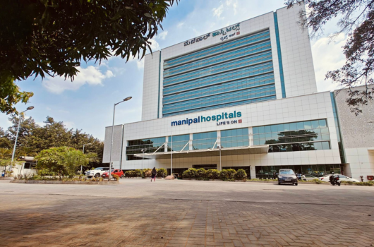
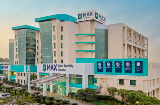
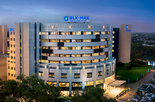
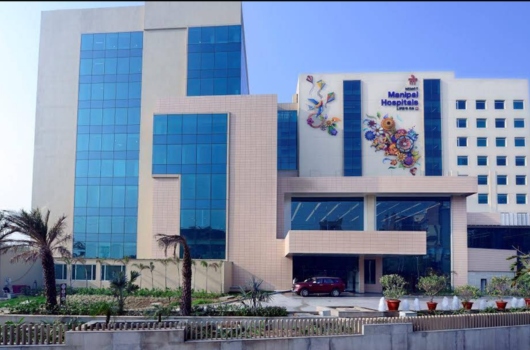
Leading hospitals for expanded endoscopic endonasal surgery include:


.png)
.png)

.png)
.png)
These hospitals follow international guidelines for care and are equipped to support patients from various countries, from diagnosis and treatment to follow-up care after returning home.
The average cost of expanded endoscopic endonasal (EEEA) surgery ranges from $3,000 to $8,500 in India and around $25,000 in Thailand. The total cost depends on factors such as tumour type and size, surgical complexity, hospital facilities, and individual patient requirements. Before reviewing detailed treatment-wise costs, it is important to understand the main factors influencing EEEA surgery expenses.
This breakdown helps patients plan for treatment expenses and understand why costs may vary between hospitals and regions.
Note: India has become a preferred destination for advanced treatment, offering world-class care at a fraction of the international cost. Patients benefit from expert doctors, modern medical technology, and affordable access to high-quality generic medicines, making treatment both effective and economical.
Note: Thailand has established itself as a premium destination, offering world-class hospitals, advanced technology, and internationally trained specialists. Patients choose Thailand not only for its high-quality medical care but also for its holistic approach, combining clinical excellence with exceptional comfort and service standards.
The above figures are approximate and can vary based on the hospital, location, and individual patient requirements. Always consult the healthcare provider for the most accurate and up-to-date pricing.
The currency conversion rates in the table above are based on data from March 2026.
For a detailed cost estimate and guidance on treatment options, patients can contact myheco to connect with leading hospitals.
A retrospective study of 29 patients who underwent the Expanded Endoscopic Endonasal Surgery (EES) for craniopharyngiomas over five years showed promising outcomes. Visual improvement or stability was achieved in 92.3% of patients, reflecting the high success of the procedure in preserving or enhancing vision. The gross total resection rate was 62%, while near-total resection (over 95% tumour removal) was achieved in 86.2% of cases.
Overall, EES offers a high success rate for tumour removal and visual preservation, making it a valuable approach for craniopharyngiomas, though endocrine and postoperative complications remain important considerations.
Success rates can vary depending on tumour type, surgical complexity, and individual patient factors.
For the expanded endoscopic endonasal approach (EEEA), success is measured by:
Leading hospitals follow a multidisciplinary, patient-focused approach for expanded endonasal procedures, combining precise diagnostics, advanced endoscopic techniques, and comprehensive postoperative care. Their approach includes:
This integrated strategy ensures safe tumour removal, preservation of critical functions, and optimal recovery, providing patients with the best possible outcomes after expanded endoscopic endonasal surgery.
From connecting patients with leading specialists to arranging online consultations and personalised treatment plans, myheco ensures a seamless and well-coordinated medical journey for international patients. With guidance on travel, hospital selection, and post-treatment care, patients can focus on recovery while myheco manages all logistics and support.
Choosing myheco means expert care, faster access, and comprehensive support throughout your treatment journey.
Note: Myheco does not provide medical advice.
✅ Share your medical reports
✅ Receive personalised treatment plans from leading hospitals
✅ Choose the option that suits you best
✅ Let us handle the arrangements

Patients with tumours or lesions at the skull base, pituitary gland, or upper spine, such as pituitary adenomas, craniopharyngiomas, meningiomas, or chordomas, may be suitable. Your neurosurgeon and ENT specialist will assess you before recommending this minimally invasive approach. Surgical eligibility varies from person to person. Always consult a qualified neurosurgeon or ENT specialist to confirm if this procedure is suitable for you.
Potential risks include cerebrospinal fluid (CSF) leaks, bleeding, infection, hormonal imbalances, and vision changes. With careful planning, high-definition imaging, and intraoperative navigation, most complications can be minimised. The risks and outcomes mentioned above are general. Your doctor will explain specific risks and precautions based on your individual case.
Hospital stay usually ranges from 3 to 7 days, depending on tumour size and complexity. Full recovery, including hormonal stabilisation, vision monitoring, and rehabilitation, may take several weeks to a few months. Recovery time varies from patient to patient. Follow your doctor’s advice for a personalised recovery plan.
Surgeons use neuronavigation and intraoperative monitoring to protect the optic nerves, pituitary gland, and cranial nerves. Most patients maintain normal vision, hormone function, and neurological health, though temporary or, rarely, permanent changes may occur. Surgical outcomes may differ based on tumour location and complexity. Discuss your specific case with your treating surgeon.
Yes. Depending on tumour type and extent, surgery may be followed by radiotherapy, radiosurgery, or targeted medical therapy to reduce the risk of recurrence. The choice of additional treatments, such as radiotherapy or medical therapy, depends on each patient’s diagnosis and should be discussed with your medical team.
Myheco connects patients with experienced skull base surgeons, arranges online consultations, helps with travel and visa logistics, ensures cost transparency, and supports patients at every stage, from preoperative planning to postoperative care and rehabilitation.
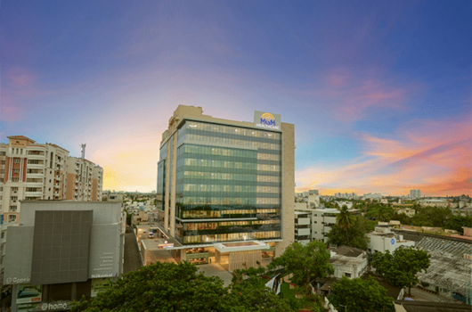
India’s leading hospitals for Expanded Endoscopic Endonasal Approach (EEEA) and advanced skull base surgery include Manipal Hospital, Apollo Hospital, and Fortis Hospital in Bangalore; Max Hospital, BLK-Max Super Speciality Hospital, Manipal Hospital Dwarka, Medanta – The Medicity, IBS (Institute of Brain and Spine), Apollo Indraprastha Hospital, and Fortis Hospital in Delhi; Nanavati Max Super Speciality Hospital, Apollo Hospital, and Fortis Hospital in Mumbai; Apollo Proton Cancer Centre (APCC), Apollo Hospital, SIMS Hospital, MGM Healthcare, and Rela Hospital in Chennai; and Apollo Hospital in Hyderabad. These centres are recognised for specialised endoscopic skull base surgery programs, high-definition endoscopic systems, neuronavigation, and multidisciplinary ENT-neurosurgery collaboration for precise tumour removal.
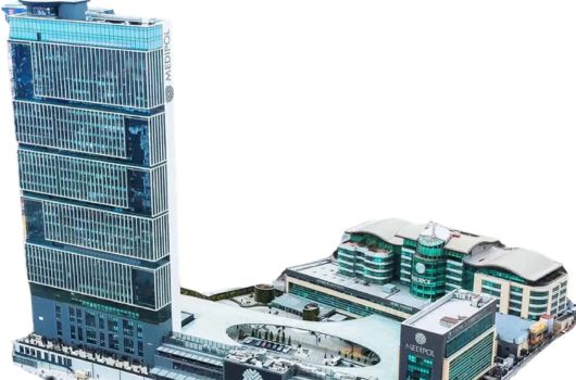
In Thailand, Samitivej Sukhumvit Hospital in Bangkok is internationally recognised for advanced minimally invasive skull base surgery, expanded endoscopic endonasal procedures, and multidisciplinary care. The hospital also provides dedicated international patient support services for patients travelling for treatment.


DISCLAIMER: THIS WEBSITE DOES NOT PROVIDE MEDICAL ADVICE
The information, including but not limited to, text, graphics, images and other material contained on this website are for informational purposes only. No material or content on this site is intended to be a substitute for professional medical advice, diagnosis, or treatment. Always seek the advice of your physician or other qualified health care provider with any questions you may have regarding a medical condition or treatment and before undertaking a new health care regimen, and never disregard professional medical advice or delay in seeking it because of something you have read on this website - www.myheco.com

.png)